Divides all cells (or alive organisms) into two types: prokaryotes And eukaryotes. Prokaryotes are nuclear-free cells or organisms, which include viruses, prokaryotic bacteria and blue-green algae, in which the cell consists directly of the cytoplasm, in which one chromosome is located - DNA molecule(sometimes RNA).
Eukaryotic cells have a core containing nucleoproteins (histone protein + DNA complex), as well as others organoids. Eukaryotes include the majority of modern unicellular and multicellular living organisms known to science (including plants).
The structure of eukaryotic granoids.
|
Organoid name |
Organoid structure |
Functions of the organoid |
|---|---|---|
|
Cytoplasm |
The internal environment of a cell in which the nucleus and other organelles are located. It has a semi-liquid, fine-grained structure. |
|
|
Ribosomes |
Small organoids of spherical or ellipsoidal shape with a diameter of 15 to 30 nanometers. |
They provide the process of synthesis of protein molecules and their assembly from amino acids. |
|
Mitochondria |
Organelles that have a wide variety of shapes - from spherical to filamentous. Inside the mitochondria there are folds from 0.2 to 0.7 µm. The outer shell of mitochondria has a double-membrane structure. The outer membrane is smooth, and on the inner there are cross-shaped outgrowths with respiratory enzymes. |
|
|
Endoplasmic reticulum (ER) |
A system of membranes in the cytoplasm that forms channels and cavities. There are two types: granular, which has ribosomes, and smooth. |
|
|
Plastids(organelles characteristic only of plant cells) are of three types: |
Double membrane organelles |
|
|
Leukoplasts |
Colorless plastids that are found in tubers, roots and bulbs of plants. |
They are an additional reservoir for storing nutrients. |
|
Chloroplasts |
Oval shaped organelles with green color. They are separated from the cytoplasm by two three-layer membranes. Chloroplasts contain chlorophyll. |
They convert organic substances from inorganic ones using solar energy. |
|
Chromoplasts |
Organelles, yellow to brown in color, in which carotene accumulates. |
Promote the appearance of yellow, orange and red colored parts in plants. |
|
Lysosomes |
Organelles are round in shape with a diameter of about 1 micron, having a membrane on the surface and a complex of enzymes inside. |
Digestive function. They digest nutrient particles and eliminate dead parts of the cell. |
|
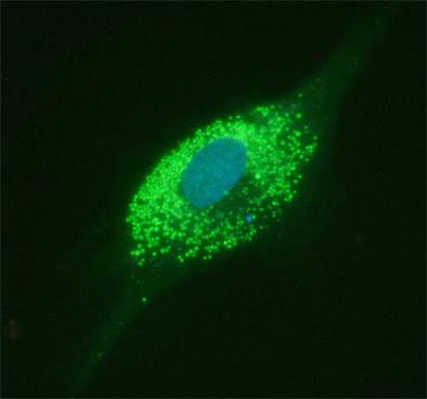
Golgi complex |
May be of different shapes. Consists of cavities delimited by membranes. Tubular formations with bubbles at the ends extend from the cavities. |
|
|
Cell center |
It consists of a centrosphere (a dense section of the cytoplasm) and centrioles - two small bodies. |
Performs an important function for cell division. |
|
Cellular inclusions |
Carbohydrates, fats and proteins, which are non-permanent components of the cell. |
Spare nutrients that are used for cell functioning. |
|
Organoids of movement |
Flagella and cilia (outgrowths and cells), myofibrils (thread-like formations) and pseudopodia (or pseudopods). |
They perform a motor function and also provide the process of muscle contraction. |
Cell nucleus is the main and most complex organelle of the cell, so we will consider it
Cell– an elementary unit of a living system. Specific functions in a cell are distributed between organoids– intracellular structures. Despite the variety of forms, cells different types have striking similarities in their main structural features.
Cell theory
As microscopes improved, new information appeared about the cellular structure of plant and animal organisms.
With the advent of physical and chemical methods The study revealed an amazing unity in the structure of cells of different organisms, and proved the inextricable connection between their structure and function.
Basic principles of cell theory
The cell is the basic unit of structure and development of all living organisms. The cells of all single- and multicellular organisms are similar in their structure, chemical composition, basic manifestations of life activity and metabolism. Cells reproduce by division. In multicellular organisms, cells are specialized in their functions and form tissues. Organs are made up of tissues.
To confirm some of the above provisions of the cell theory, let us call common features, characteristic of animal and plant cells.
Common characteristics of plant and animal cells
Unity of structural systems - cytoplasm and nucleus. The similarity of metabolic and energy processes. Unity of the principle of hereditary code. Universal membrane structure. Unity of chemical composition. Similarities in the process of cell division.
Table Features plant and animal cells
Signs | plant cell | animal cell |
Plastids | Chloroplasts, chromoplasts, leucoplasts | Absent |
Nutrition method | Autotrophic (phototrophic, chemotrophic). | Heterotrophic (saprotrophic, chemotrophic). |
ATP synthesis | In chloroplasts, mitochondria. | In mitochondria. |
ATP breakdown | In chloroplasts and all parts of the cell where energy is required. |
|
Cell center | In lower plants. | In all cells. |
Cellulose cell wall | Located outside the cell membrane. | Absent. |
Inclusion | Spare nutrients in the form of grains of starch, protein, drops of oil; in vacuoles with cell sap; salt crystals. | Spare nutrients in the form of grains and drops (proteins, fats, carbohydrate glycogen); end products of metabolism, salt crystals; pigments. |
Large cavities filled with cell sap - an aqueous solution of various substances that are reserve or final products. Osmotic reservoirs of the cell. | Contractile, digestive, excretory vacuoles. Usually small. |
The importance of theory: it proves the unity of origin of all living organisms on Earth.
Cellular structures
Figure Scheme of the structure of animal and plant cells

Organelles | Structure | Functions |
Cytoplasm | Located between the plasma membrane and the nucleus, it includes various organelles. The space between the organelles is filled with cytosol - a viscous aqueous solution of various salts and organic substances, permeated with a system of protein threads - the cytoskeleton. | Most of the chemical and physiological processes of the cell take place in the cytoplasm. Cytoplasm unites all cellular structures into a single system and ensures the relationship between the exchange of substances and energy between the organelles of the cell. |
Outer cell membrane | An ultramicroscopic film consisting of two monomolecular layers of protein and a bimolecular layer of lipids located between them. The integrity of the lipid layer can be interrupted by protein molecules - “pores”. | Isolates the cell from environment, has selective permeability, regulates the process of substances entering the cell; ensures the exchange of substances and energy with the external environment, promotes the connection of cells in tissue, participates in pinocytosis and phagocytosis; regulates water balance cells and removes waste products from it. |
Endoplasmic reticulum (ER) | Ultramicroscopic system of membranes forming tubes, tubules, cisterns, vesicles. The structure of the membranes is universal (as well as the outer one), the entire network is united into a single whole with the outer membrane of the nuclear membrane and the outer cellular membrane. The granular ES carries ribosomes, while the smooth one lacks them. | Provides transport of substances both within the cell and between neighboring cells. Divides the cell into separate sections in which various physiological processes occur simultaneously and chemical reactions. Granular ES is involved in protein synthesis. Complex protein molecules are formed in ES channels, fats are synthesized, and ATP is transported. |
Ribosomes | Small spherical organelles consisting of rRNA and protein. | Proteins are synthesized on ribosomes. |
Golgi apparatus | Microscopic single-membrane organelles, consisting of a stack of flat cisterns, along the edges of which tubes branch off, separating small vesicles. | In the general system of membranes of any cells, it is the most mobile and changing organelle. The cisterns accumulate decomposition synthesis products and substances that enter the cell, as well as substances that are removed from the cell. Packed in vesicles, they enter the cytoplasm: some are used, while others are excreted. |
Lysosomes | Microscopic single-membrane organelles of round shape. Their number depends on the vital activity of the cell and its physiological state. Lysosomes contain lysing (dissolving) enzymes synthesized on ribosomes. | Digestion of food that enters an animal cell during phagocytosis and pinocytosis. Protective function. In the cells of any organisms, autolysis (self-dissolution of organelles) occurs; especially under conditions of food or oxygen starvation, the tail of animals dissolves. In plants, organelles dissolve during the formation of cork tissue of wood vessels. |
Conclusions from the lecture
An important achievement biological science is the formation of ideas about the structure and vital activity of the cell as a structural and functional unit of the body. The science that studies the living cell in all its manifestations is called cytology. The first stages of the development of cytology as a field of scientific knowledge were associated with the works of R. Hooke, A. Leeuwenhoek, T. Schwann, M. Schleiden, R. Virchow, K. Baer. The result of their activity was the formulation and development of the basic principles of cell theory. A variety of cellular structures are directly involved in the vital processes of a cell. Cytoplasm ensures the activity of all cellular structures as a single system. The cytoplasmic membrane ensures the passage selectivity of substances in the cell and protects it from external environment. The ES ensures the transport of substances both within the cell and between neighboring cells. In the tanks of the Golgi Apparatus, products of the synthesis and breakdown of substances entering the cell, as well as substances that are removed from the cell, accumulate. Lysosomes break down substances that enter the cell.
Questions for self-control
Using knowledge of cell theory, prove the unity of the origin of life on Earth. What are the similarities and differences in the structure of plant and animal cells? How is the structure of the cell membrane related to its functions? How does active absorption of substances into cells occur? What is the connection between ribosomes and ES? What are the structure and functions of lysosomes in a cell?
Cellular structures: mitochondria, plastids, organelles of movement, inclusions. Core
Table Cell organelles, their structure and functions
Organelles | Structure | Functions |
Mitochondria | Microscopic organelles with a double-membrane structure. The outer membrane is smooth, the inner one forms various shapes outgrowths - cristae. The mitochondrial matrix (a semi-liquid substance) contains enzymes, ribosomes, DNA, and RNA. | The universal organelle is the respiratory and energy center. During the oxygen (oxidative) stage in the matrix, with the help of enzymes, organic substances are broken down with the release of energy, which goes to the synthesis of ATP on (cristae). |
Leukoplasts | Microscopic organelles with a double-membrane structure. The inner membrane forms 2–3 outgrowths. The shape is round. Colorless. | Characteristic of plant cells. They serve as a site for the deposition of reserve nutrients, mainly starch grains. In the light, their structure becomes more complex, and they transform into chloroplasts. Formed from proplastids. |
Chloroplasts | Microscopic organelles with a double-membrane structure. The outer membrane is smooth. The inner membrane forms a system of two-layer plates - stromal thylakoids and granal thylakoids. Pigments - chlorophyll and carotenoids - are concentrated in the membranes of thylakoid granules between layers of protein and lipid molecules. The protein-lipid matrix contains its own ribosomes, DNA, and RNA. | Characteristic of plant cells are photosynthesis organelles that are capable of creating organic substances - carbohydrates and free oxygen - from inorganic substances (CO2 and H2O) in the presence of light energy and the pigment chlorophyll. Synthesis of own proteins. They can be formed from plastids or leucoplasts, and in the fall they turn into chloroplasts (red and orange fruits, red and yellow leaves). |
Chromoplasts | Microscopic organelles with a double-membrane structure. Chromoplasts themselves have a spherical shape, and those formed from chloroplasts take the form of caratinodon crystals, typical for this type of plant. Color: red, orange, yellow. | Characteristic of plant cells. They give flower petals a color that is attractive to pollinating insects. IN autumn leaves and ripe fruits separated from plants contain crystalline carotenoids - end products of metabolism. |
Cell center | Ultramicroscopic organelle of non-membrane structure. Consists of two centrioles. Each has a cylindrical shape, the walls are formed by nine triplets of tubes, and in the middle there is a homogeneous substance. The centrioles are located perpendicular to each other. | Takes part in the division of cells of animals and lower plants. At the beginning of division (in prophase), the centrioles diverge to different poles of the cell. The spindle strands extend from the centrioles to the centromeres of the chromosomes. In anaphase, these threads attract chromatids to the poles. After the end of division, the centrioles remain in the daughter cells. They double and form a cell center. |
Cellular inclusions (non-permanent structures) | Dense, granular inclusions with a membrane (for example, vacuoles). | |
Organoids of movement | Cilia are numerous cytoplasmic projections on the surface of the membrane. | Removal of dust particles (ciliated epithelium of the upper respiratory tract), movement (single-celled organisms). |
Flagella are single cytoplasmic projections on the cell surface. | Movement (spermatozoa, zoospores, single-celled organisms). |
|
False legs (pseudopodia) are amoeboid protrusions of the cytoplasm. | Formed in animals different places cytoplasm for food capture and movement. |
|
Myofibrils are thin filaments up to 1 cm long or more. | They serve to contract the muscle fibers along which they are located. |
|
Cytoplasm, which carries out stream and circular movement. | Movement of cell organelles in relation to (during photosynthesis), heat, chemical irritant. |
Figure Scheme of composition and functions of cellular inclusions

Phagocytosis– capture of solid particles by the plasma membrane and draw them inward.
The plasma membrane forms an invagination in the form of a thin tubule into which liquid with substances dissolved in it enters. This method is called pinocenosis.
Core
All organisms that have a cellular structure without a formed nucleus are called prokaryotes. All organisms that have a cellular structure with a nucleus are called eukaryotes.
Table Nuclear structures, their structure and functions
Structures | Structure | Functions |
Nuclear envelope | Double-layer porous. The outer membrane passes into the ES membranes. It is characteristic of all animal and plant cells, except bacteria and blue-green ones, which do not have a nucleus. | Separates the nucleus from the cytoplasm. Regulates the transport of substances from the nucleus to the cytoplasm (RNA and ribosomal subunits) and from the cytoplasm to the nucleus (proteins, fat, carbohydrates, ATP, water, ions). |
Chromosomes (chromatin) | In an interphase cell, chromatin has the form of fine-grained thread-like structures consisting of DNA molecules and a protein sheath. In dividing cells, chromatin structures spiral and form chromosomes. A chromosome consists of two chromatids, and after nuclear division it becomes single chromatid. By the beginning of the next division, a second chromatid is completed on each chromosome. Chromosomes have a primary constriction on which the centromere is located; the constriction divides the chromosome into two arms of equal or different lengths. Nucleolar chromosomes have a secondary constriction. | Chromatin structures are carriers of DNA. DNA consists of sections - genes that carry hereditary information and are transmitted from ancestors to descendants through germ cells. The totality of chromosomes, and, consequently, the genes of the germ cells of the parents, is transmitted to children, which ensures the stability of the characteristics characteristic of a given population or species. DNA and RNA are synthesized in chromosomes, which serves as a necessary factor in the transmission of hereditary information during cell division and the construction of protein molecules. |
A spherical body resembling a ball of thread. Consists of protein and RNA. Formed on the secondary constriction of the nucleolar chromosome. It breaks down when cells divide. | Formation of ribosome halves from rRNA and protein. The halves (subunits) of ribosomes enter the cytoplasm through pores in the nuclear envelope and combine to form ribosomes. |
|
Nuclear juice (karyolymph) | A semi-liquid substance representing a colloidal solution of proteins, nucleic acids, carbohydrates, and mineral salts. The reaction is sour. | Participates in the transport of substances and nuclear structures, fills the space between nuclear structures; During cell division it mixes with the cytoplasm. |
Figure Scheme of the structure of the cell nucleus
Functions of the cell nucleus:
- regulation of metabolic processes in the cell; storage of hereditary information and its reproduction; RNA synthesis; ribosome assembly.
Conclusions from the lecture
In mitochondria, organic substances are broken down and energy is released, which is used for the synthesis of ATP. Plastids play an important role in ensuring the vital processes of the plant cell. Organelles of movement include cellular structures: cilia, flagella, myofibrils. All cellular organisms are divided into prokaryotes (without a nucleus) and eukaryotes (with a nucleus). The nucleus is a structural and functional center that coordinates its metabolism, directing the processes of self-reproduction and storage of hereditary information.
Questions for self-control
Why are mitochondria figuratively called the “power stations” of the cell? What cell structures contribute to its movement? What are cellular inclusions? What is their role? What are the functions of the nucleus in a cell?
Organic substances in the cell (carbohydrates, proteins, lipids, nucleic acids, ATP, vitamins, etc.)
Biological polymers– organic compounds that make up the cells of living organisms. Polymer is a multi-link chain of simple substances – monomers (n ÷ 10 thousand – 100 thousand monomers)

The properties of biopolymers depend on the structure of their molecules, on the number and variety of monomer units.
If the monomers are different, then their repeated alternations in the chain create a regular polymer.
…A – A – B – A – A – B... regular
…A – A – B – B – A – B – A... irregular
Carbohydrates
General formula Сn(H2O)m
Carbohydrates play the role of energy substances in the human body. The most important of them are - sucrose, glucose, fructose, and starch. They are quickly absorbed ("burned") in the body. The exception is cellulose(cellulose), which is especially abundant in plant foods. It is practically not absorbed by the body, but has great importance: Acts as ballast and aids digestion by mechanically cleansing the mucous membranes of the stomach and intestines. There are a lot of carbohydrates in potatoes and vegetables, cereals, pasta, fruits and bread.
Glucose, ribose, fructose, deoxyribose - monosaccharides
Sucrose - disaccharides
Starch, glycogen, cellulose - polysaccharides
Finding in nature: in plants, fruits, pollen, vegetables (garlic, beets), potatoes, rice, corn, wheat grain, wood...
Their functions:
- energy: oxidation to CO2 and H2O releases energy; excess energy is stored in liver and muscle cells in the form of glycogen; construction: in a plant cell - a strong base of cell walls (cellulose); structural: part of the intercellular substance of the skin, cartilage tendons; recognition by other cells: as part of cell membranes, if separated liver cells are mixed with kidney cells, they will independently separate into two groups due to the interaction of cells of the same type.
Lipids (lipoids, fats)
Lipids include various fats, fat-like substances, phospholipids... All of them are insoluble in water, but soluble in chloroform, ether...
Finding in nature: in animal and human cells in the cell membrane; between the cells is the subcutaneous layer of fat.
Functions:
- thermal insulation (in whales, pinnipeds...); storage nutrient; energy: energy is released during the hydrolysis of fats; structural: some lipids serve integral part cell membranes.
Fats also serve as a source of energy for the human body. The body stores them “in reserve” and they serve as a long-term energy source. In addition, fats have low thermal conductivity and protect the body from hypothermia. It is not surprising that in the traditional diet northern peoples so many animal fats. For people engaged in heavy physical labor, it is also easiest (although not always healthier) to compensate for the energy expended with fatty foods. Fats are part of cell walls, intracellular formations, and nervous tissue. Another function of fats is to supply fat-soluble vitamins and other biologically active substances to the body tissues.
Squirrels
Figure 1.2.1. Protein molecule


If in R we replace one more H with the amino group NH2, we get the amino acid:

Proteins are biopolymers whose monomers are amino acids.
The formation of linear protein molecules occurs as a result of reactions of amino acids with each other.

Sources of proteins can be not only animal products (meat, fish, eggs, cottage cheese), but also plant products, for example, legumes (beans, peas, soybeans, peanuts, which contain up to 22–23% proteins by weight), nuts and mushrooms . However, cheese contains the most protein (up to 25%), meat products(in pork 8–15%, lamb 16–17%, beef 16–20%), in poultry (21%), fish (13–21%), eggs (13%), cottage cheese (14%). Milk contains 3% proteins, and bread 7–8%. Among cereals, the champion in proteins is buckwheat (13% of proteins in dry cereals), which is why it is recommended for dietary nutrition. To avoid “excesses” and at the same time ensure the normal functioning of the body, it is necessary, first of all, to give a person a complete set of proteins with food. If there is not enough protein in the diet, an adult feels a loss of strength, his performance decreases, and his body is less resistant to infections and colds. As for children, if they have inadequate protein nutrition, they are greatly behind in development: children grow, and proteins are the main “building material” of nature. Every cell of a living organism contains proteins. Human muscles, skin, hair, and nails consist mainly of proteins. Moreover, proteins are the basis of life; they participate in metabolism and ensure the reproduction of living organisms.
Structure:
- primary structure – linear, with alternating amino acids; secondary - in the form of a spiral with weak bonds between the turns (hydrogen); tertiary - a spiral rolled into a ball; quaternary - when combining several chains that differ in primary structure.
With radiation, high temperatures, extreme pH values, in alcohol, acetone, the protein is destroyed - a denaturation reaction.
Table 1.2.1. Protein structure
| Primary structure– a specific sequence of α-amino acid residues in a polypeptide chain |
Secondary structure– conformation of a polypeptide chain, secured by many hydrogen bonds between groups N-H and C=O. One of the models of secondary structure is an α-helix due to cooperative intramolecular H-bonds. Another model is the b-form (“folded sheet”), in which interchain (intermolecular) H-bonds predominate |
|
| Tertiary structure- the shape of a twisted helix in space, formed mainly due to disulfide bridges - S-S-, hydrogen bonds, hydrophobic and ionic interactions |
| Quaternary structure– aggregates of several protein macromolecules (protein complexes), formed through the interaction of different polypeptide chains |


Functions:
- construction: proteins are an essential component of all cellular structures; structural: proteins in combination with DNA make up the body of chromosomes, and with RNA – the body of ribosomes; enzymatic: chemical catalyst. reactions are performed by any enzyme - a protein, but a very specific one; transport: transfer of O2, hormones in the body of animals and humans; regulatory: proteins can perform a regulatory function if they are hormones. For example, insulin (a hormone that supports the functioning of the pancreas) activates the uptake of glucose molecules by cells and their breakdown or storage inside the cell. With a lack of insulin, glucose accumulates in the blood, developing diabetes; protective: when foreign bodies enter the body, protective proteins are produced - antibodies, which bind to foreign bodies, combine and suppress their vital functions. This mechanism of resistance of the body is called immunity; energy: with a lack of carbohydrates and fats, amino acid molecules can be oxidized.
Adenosine triphosphoric acid (ATP)– a universal carrier and main energy accumulator in living maples, which is necessary for the synthesis of organic substances, movement, heat production, nerve impulses, glows. ATP is found in all plant and animal cells.
It is a nucleotide formed by residues of a nitrogenous base (adenine), a sugar (ribose) and three phosphoric acid residues.
ATP is an unstable molecule: when the terminal phosphoric acid residue is removed. ATP is converted into ADP (adenosine diphosphoric acid), and about 30.5 kJ is released.
Figure 1.2.2. The structure of the ATP molecule

Hormones organic compounds, which may be of a protein nature (pancreatic hormones) and may be lipids (sex hormones), may be derivatives of amino acids. Hormones are produced by both animals and plants. Hormones carry out various different functions:
- regulate the content of sodium ions and water in the body; ensure puberty; anxiety and stress hormones increase the release of glucose into the blood and, therefore, determine the active use of energy; signaling hormones report the presence of food and danger; Plants have their own hormones that accelerate the ripening of fruits and attract insects.
Nucleic acids– biopolymers whose monomers are nucleotides.
Figure 1.2.3. Nucleic acid synthesis

Figure 1.2.4. Schematic structure of DNA (ellipses indicate hydrogen bonds)

The DNA molecule is a structure consisting of two strands, which are connected to each other along their entire length by hydrogen bonds. (Fig. 1.2.4)
Figure 1.2.5. Section of a DNA molecule

A feature of the DNA structure is that opposite the nitrogenous base A in one chain lies the nitrogenous base T in the other chain, and opposite the nitrogenous base G is always the nitrogenous base C. The above can be shown in the form of a diagram:

These base pairs are called complementary bases (complementary to each other). DNA strands in which the bases are located complementary to each other are called complementary strands. In Fig. Figure 1.2.5 shows two strands of DNA that are connected by complementary regions.
The order of nucleotides in DNA molecules determines the order of amino acids in linear protein molecules.
Table Comparative characteristics DNA and RNA
Signs of comparison | ||
Location in the cage | Nucleus, mitochondria, chloroplasts | Nucleus, ribosomes, cytoplasm, mitochondria, chloroplasts |
Location in the nucleus | Chromosomes | |
Structure of a macromolecule | Double unbranched linear polymer, coiled in a right-handed helix | Single polynucleotide chain |
Composition of nucotides | Nitrogen base (adenine, guanine, thymine, cytosine); deoxyribose (carbohydrate); phosphoric acid residue | Nitrogen base (adenine, guanine, uracil, cytosine); ribose (carbohydrate); phosphoric acid residue |
Chemical basis chromosomal genetic material (gene); DNA and RNA synthesis, information about protein structure | Information (mRNA) transmits the code of hereditary information about the primary structure of the protein molecule; ribosomal (rRNA) is part of ribosomes; transport (tRNA) carries amino acids to ribosomes. |
Vitamins
Back at the end of the 19th century, scientists discovered that the terrible beri-beri disease, in which damage occurs nervous system, is caused by a lack of some special substance in food. In 1912, Polish researcher Kazimierz Funk (1884–1967) isolated a substance from rice bran and called it vitamin (from the Latin vita - “life”). That's what they call it chemical compounds, which are required for the normal functioning of the body in very small quantities. The body “does not know how” to synthesize vitamins on its own. Therefore, it is very important to replenish the body with vitamin-containing foods. A lack of vitamins in the body is the cause of a serious disease - vitamin deficiency.
A healthy person under normal living conditions should try to fully cover his need for vitamins through a varied and nutritious diet. You should turn to pharmaceutical preparations containing vitamins in cases where you experience a permanent or seasonal (autumn, spring) deficiency of vitamins, as well as under severe stress. Unsystematic amateur “eating” of vitamin pills can cause unpleasant consequences in the form of hypervitaminosis, when even required amount Vitamins are not absorbed but excreted by the body.
Vitamins
Back in the late 19th century, scientists discovered that the terrible beriberi disease, which damages the nervous system, is caused by a lack of some special substance in food. In 1912, Polish researcher Kazimierz Funk (1884–1967) isolated such a substance from rice bran and called it a vitamin (from the Latin vita - “life”). About 25 vitamins have now been well studied. Chemical composition and their names are very complex, so they were assigned alphabetic symbols. It is customary to divide all vitamins into two large groups: water-soluble And fat-soluble.
The main water-soluble vitamins are:
1. B1 – thiamine, first found in white cabbage; then it was also found in some cereals, raw fish, yeast and sprouted wheat. This vitamin regulates metabolism, nervous activity and is responsible for the condition of the cardiovascular system. The lack of B1 in food causes beriberi, a severe joint disease associated with damage to the nervous system, heart and blood vessels. Beriberi is common in those regions of Southeast Asia where the population eats a poor and monotonous diet, mainly only refined rice, which contains almost no vitamin B1. The body's daily need for vitamin B1 is 1.5–2.0 mg.
2. B2 – riboflavin. Regulates metabolism, increases visual acuity, improves liver and nervous system function, as well as skin condition. Sources of vitamin B2 are yeast, meat, fish, liver and other offal (kidneys, heart, tongue), egg yolk, dairy products, legumes and many cereals. The body's daily need for vitamin B2 is 2.0–2.5 mg;
3. RR – a nicotinic acid(niacin) regulates cellular respiration and cardiac activity. Sources of vitamin PP include yeast, meat and dairy products, and grain crops. In addition, it is one of the few vitamins that can be produced in the human body. Vitamin PP is formed from tryptophan, an amino acid that is part of proteins supplied with food. The body's daily need for vitamin PP is 15–20 mg;
4. B6 – pyridoxine, participates in metabolic processes, is necessary for the absorption of amino acids and for the synthesis of vitamin PP from tryptophan. The body's daily need for vitamin B6 is 2 mg;
5. BC – folacin, folic acid and its derivatives, regulate hematopoiesis and fat metabolism. Contained in liver, yeast, and many vegetables (parsley, spinach, and lettuce). The body's daily need for vitamin BC is 2.0–2.5 mg.
6. B12 – cyanocobalamin. Prevents anemia. Present in beef and pork liver, rabbit and chicken meat, eggs, fish, milk. The body's daily requirement for vitamin B12 is 3 mg.
7. C – ascorbic acid, protects against scurvy, improves immunity. Sources of this vitamin in the diet are fresh and canned vegetables, fruits, and berries. Rose hips, currants, parsley, dill are especially rich in ascorbic acid, and among the wild ones there are nettles, sorrel, and wild garlic. Ascorbic acid is unstable: in air it easily oxidizes to dehydroascorbic acid, which does not have vitamin properties. This must be taken into account when cooking vegetables and fruits. The body's daily need for vitamin C is 75–100 mg.
8. R – routine(bioflavonoid) is a vascular strengthening agent, is active together with vitamin C. There is especially a lot of it in currants, rose hips, chokeberry(chokeberry), citrus and green tea. The body's daily need for vitamin P is 25–50 mg.
Among the fat-soluble vitamins, the most important are:
1. A – retinol and its derivatives, improves the condition of the skin and mucous membranes of the eyes, increases immunity, and most importantly, ensures visual acuity in the twilight. With a lack of vitamin A, “night blindness” occurs (a person has difficulty seeing in evening time). Retinol is found in milk, butter, cheese, fish oil, and can also be synthesized in the human liver from provitamin A - carotene, the source of which is carrots, tomatoes and sea buckthorn. The body's daily need for vitamin A is 1.5 - 2.0 mg (or 6 mg of carotene);
2. D – ergocalciferol, has an antirachitic effect and helps the absorption of calcium. It is absolutely necessary for a growing body during the formation and development of bones and teeth. Vitamin D is found in fish oil, caviar, butter, eggs, and milk. In addition, it is formed in the body under the influence sun rays. The body's daily requirement for vitamin D is 0.01 mg.
3. E – tocopherol, affects the functions of the gonads and promotes the normal course of pregnancy, promotes the absorption of fat-soluble vitamins, and participates in metabolism. Contained in vegetable oil, buckwheat, legumes. The body's daily requirement for vitamin E is 12–15 mg.
4. K – antihemorrhagic factor, regulates blood clotting, prevents bleeding. Sources of this vitamin include potatoes, cabbage, pumpkin, spinach, sorrel, and liver. The body's daily requirement for vitamin K is 0.2–0.3 mg.
Conclusions from the lecture
The main organic substances in the cell include proteins, carbohydrates, fats, nucleic acids and ATP. Carbohydrates play the role of energy substances in the life of plants, animals, fungi and microorganisms. Fats are the main structural component of cell membranes and a source of energy. They undergo complex transformations in the cell. Proteins are biological polymers, the monomers of which are 20 essential amino acids, and perform a number of important functions in the cell. Construction: proteins are an essential component of all cellular structures; structural: proteins in combination with DNA make up the body of chromosomes, and with RNA – the body of ribosomes; enzymatic: chemical catalyst. reactions – specific enzyme – protein; transport: transfer of O2, hormones in the body of animals and humans; regulatory: (hormones) part of hormones - proteins, for example insulin - a hormone that supports glands, activates the uptake of glucose molecules by cells and their breakdown or storage inside the cell. With a lack of insulin, glucose accumulates in the blood, developing diabetes; protective: when foreign bodies enter the body, protective proteins are produced - antibodies, which bind to foreign bodies, combine and suppress their vital activity. This mechanism of resistance of the body is called immunity; energy: with a lack of carbohydrates and fats, amino acid molecules can oxidize. DNA - molecules of heredity, consist of monomers - nucleotides. DNA and RNA nucleotides have similarities and differences in structure and perform different functions. The great importance of vitamins for organisms has been revealed.
Questions for self-control
What carbohydrates are characteristic of a plant cell and an animal cell? Specify the functions of carbohydrates. Describe the structure of protein molecules in connection with their functions in the cell. What is the primary, secondary, tertiary and quaternary structure of a protein molecule? What is special about the structure of the DNA molecule? What components make up nucleotides? What functions do DNA and RNA perform?
Based on materials from the site http://umka. *****
The elementary and functional unit of all life on our planet is the cell. In this article you will learn in detail about its structure, the functions of organelles, and also find the answer to the question: “How is the structure of plant and animal cells different?”
Cell structure
The science that studies the structure of the cell and its functions is called cytology. Despite their small size, these parts of the body have a complex structure. Inside is a semi-liquid substance called cytoplasm. All vital processes take place here and the component parts - organelles - are located. You can learn about their features below.
Core
The most important part is the core. It is separated from the cytoplasm by a shell, which consists of two membranes. They have pores so that substances can pass from the nucleus into the cytoplasm and vice versa. Inside there is nuclear juice (karyoplasm), in which the nucleolus and chromatin are located.

Rice. 1. Structure of the nucleus.
It is the nucleus that controls the life of the cell and stores genetic information.
The functions of the internal contents of the nucleus are the synthesis of protein and RNA. From them special organelles are formed - ribosomes.
Ribosomes
They are located around the endoplasmic reticulum, making its surface rough. Sometimes ribosomes are freely located in the cytoplasm. Their functions include protein biosynthesis.
TOP 4 articleswho are reading along with this
Endoplasmic reticulum
EPS can have a rough or smooth surface. The rough surface is formed due to the presence of ribosomes on it.
The functions of the EPS include protein synthesis and internal transport of substances. Part of the formed proteins, carbohydrates and fats enters special storage containers through the channels of the endoplasmic reticulum. These cavities are called the Golgi apparatus; they are presented in the form of stacks of “cisterns”, which are separated from the cytoplasm by a membrane.
Golgi apparatus
Most often located near the nucleus. Its functions include protein conversion and the formation of lysosomes. This complex stores substances that were synthesized by the cell itself for the needs of the whole organism, and will later be removed from it.
Lysosomes are presented in the form of digestive enzymes, which are enclosed by a membrane in vesicles and distributed throughout the cytoplasm.
Mitochondria
These organelles are covered with a double membrane:
- smooth - outer shell;
- cristae - an inner layer with folds and protrusions.

Rice. 2. The structure of mitochondria.
The functions of mitochondria are respiration and conversion of nutrients into energy. The cristae contain an enzyme that synthesizes ATP molecules from nutrients. This substance is universal source energy for all kinds of processes.
The cell wall separates and protects the internal contents from the external environment. It maintains shape, ensures communication with other cells, and ensures the metabolic process. The membrane consists of a double layer of lipids, between which there are proteins.
Comparative characteristics
Plant and animal cells differ from each other in their structure, size and shape. Namely:
- the cell wall of a plant organism has a dense structure due to the presence of cellulose;
- a plant cell has plastids and vacuoles;
- an animal cell has centrioles, which are important in the process of division;
- The outer membrane of an animal organism is flexible and can take on various shapes.

Rice. 3. Scheme of the structure of plant and animal cells.
The following table will help summarize knowledge about the main parts of the cellular organism:
Table "Cell structure"
|
Organoid |
Characteristic |
Functions |
|
It has a nuclear envelope, which contains nuclear sap with a nucleolus and chromatin. |
Transcription and storage of DNA. |
|
|
Plasma membrane |
It consists of two layers of lipids, which are permeated with proteins. |
Protects the contents, ensures intercellular metabolic processes, and responds to stimuli. |
|
Cytoplasm |
Semi-liquid mass containing lipids, proteins, polysaccharides, etc. |
Association and interaction of organelles. |
|
Membrane bags of two types (smooth and rough) |
Synthesis and transportation of proteins, lipids, steroids. |
|
|
Golgi apparatus |
Located near the nucleus in the form of vesicles or membrane sacs. |
Forms lysosomes and removes secretions. |
|
Ribosomes |
They have protein and RNA. |
They form protein. |
|
Lysosomes |
In the form of a bag containing enzymes. |
Digestion of nutrients and dead parts. |
|
Mitochondria |
The outside is covered with a membrane and contains cristae and numerous enzymes. |
Formation of ATP and protein. |
|
Plastids |
Covered with a membrane. They are represented by three types: chloroplasts, leucoplasts, chromoplasts. |
Photosynthesis and storage of substances. |
|
Sacs with cell sap. |
Regulate blood pressure and retain nutrients. |
|
|
Centrioles |
Has DNA, RNA, proteins, lipids, carbohydrates. |
Participates in the process of division, forming a spindle. |
What have we learned?
A living organism consists of cells that have a rather complex structure. On the outside, it is covered with a dense shell that protects the internal contents from exposure to the external environment. Inside there is a core that regulates all ongoing processes and stores the genetic code. Around the nucleus there is cytoplasm with organelles, each of which has its own characteristics and characteristics.
Test on the topic
Evaluation of the report
Average rating: 4.3. Total ratings received: 1075.
The science that studies the structure and function of cells is called cytology.
Cell- an elementary structural and functional unit of living things.
Cells, despite their small size, are very complex. The internal semi-liquid contents of the cell are called cytoplasm.
Cytoplasm is the internal environment of the cell where various processes and the components of the cell - organelles (organelles) are located.
Cell nucleus
The cell nucleus is the most important part of the cell.
The nucleus is separated from the cytoplasm by a shell consisting of two membranes. The nuclear membrane has numerous pores so that various substances can enter the nucleus from the cytoplasm and vice versa.
The internal contents of the kernel are called karyoplasma or nuclear juice. Located in the nuclear juice chromatin And nucleolus.
Chromatin is a strand of DNA. If the cell begins to divide, then the chromatin threads are tightly wound into a spiral around special proteins, like threads on a spool. Such dense formations are clearly visible under a microscope and are called chromosomes.

Core contains genetic information and controls the life of the cell.
Nucleolus is a dense round body inside the core. Typically, there are from one to seven nucleoli in the cell nucleus. They are clearly visible between cell divisions, and during division they are destroyed.

The function of the nucleoli is the synthesis of RNA and proteins, from which special organelles are formed - ribosomes.
Ribosomes participate in protein biosynthesis. In the cytoplasm, ribosomes are most often located on rough endoplasmic reticulum. Less commonly, they are freely suspended in the cytoplasm of the cell.
Endoplasmic reticulum (ER) participates in the synthesis of cell proteins and transport of substances within the cell.

A significant part of the substances synthesized by the cell (proteins, fats, carbohydrates) is not consumed immediately, but through the EPS channels enters for storage in special cavities laid in peculiar stacks, “cisterns”, and delimited from the cytoplasm by a membrane. These cavities are called Golgi apparatus (complex). Most often, the cisterns of the Golgi apparatus are located close to the cell nucleus.
Golgi apparatus takes part in the transformation of cell proteins and synthesizes lysosomes- digestive organelles of the cell.
Lysosomes They are digestive enzymes, “packed” into membrane vesicles, budded and distributed throughout the cytoplasm.
The Golgi complex also accumulates substances that the cell synthesizes for the needs of the whole organism and which are removed from the cell to the outside.

Mitochondria- energy organelles of cells. They convert nutrients into energy (ATP) and participate in cell respiration.
Mitochondria are covered with two membranes: the outer membrane is smooth, and the inner one has numerous folds and projections - cristae.

Plasma membrane
For a cell to be a single system, it is necessary that all its parts (cytoplasm, nucleus, organelles) are held together. For this purpose, in the process of evolution, it developed plasma membrane, which, surrounding each cell, separates it from the external environment. The outer membrane protects the internal contents of the cell - the cytoplasm and nucleus - from damage, maintains a constant shape of the cell, ensures communication between cells, selectively allows necessary substances into the cell and removes metabolic products from the cell.
The structure of the membrane is the same in all cells. The basis of the membrane is a double layer of lipid molecules, in which numerous protein molecules are located. Some proteins are located on the surface of the lipid layer, others penetrate both layers of lipids through and through.

Special proteins form the finest channels through which potassium, sodium, calcium ions and some other ions of small diameter can pass into or out of the cell. However, larger particles (nutrient molecules - proteins, carbohydrates, lipids) cannot pass through membrane channels and enter the cell using phagocytosis or pinocytosis:
- At the point where the food particle touches the outer membrane of the cell, an invagination is formed, and the particle enters the cell, surrounded by a membrane. This process is called phagocytosis (plant cells are covered with a dense layer of fiber (cell membrane) on top of the outer cell membrane and cannot capture substances by phagocytosis).
- Pinocytosis differs from phagocytosis only in that in this case the invagination of the outer membrane captures not solid particles, but droplets of liquid with substances dissolved in it. This is one of the main mechanisms for the penetration of substances into the cell.
An independent biosystem that possesses the basic properties of all living things. So, it can develop, reproduce, move, adapt and change. In addition, any cells are characterized by metabolism, specific structure, orderliness of structures and functions.
The science that studies cells is cytology. Its subject is the structural units of multicellular animals and plants, unicellular organisms - bacteria, protozoa and algae, consisting of just one cell.
If we talk about the general organization of the structural units of living organisms, they consist of a shell and a nucleus with a nucleolus. They also include cell organelles and cytoplasm. Today, a variety of research methods are highly developed, but the leading place is occupied by microscopy, which allows one to study the structure of cells and study its main structural elements.
What is an organoid?
Organelles (also called organelles) are permanent constituent elements of any cell that make it whole and perform certain functions. These are structures that are vital to maintaining its activities.
Organelles include the nucleus, lysosomes, endoplasmic reticulum and Golgi complex, vacuoles and vesicles, mitochondria, ribosomes, and the cell center (centrosome). This also includes structures that form the cell cytoskeleton (microtubules and microfilaments), melanosomes. The organelles of movement should be highlighted separately. These are cilia, flagella, myofibrils and pseudopods.
All these structures are interconnected and ensure coordinated cell activity. That is why the question: “What is an organoid?” - we can answer that this is a component that can be equated to an organ of a multicellular organism.
Classification of organelles
Cells differ in size and shape, as well as in their functions, but at the same time they have a similar chemical structure and a single principle of organization. At the same time, the question of what an organoid is and what structures it is is quite debatable. For example, lysosomes or vacuoles are sometimes not classified as cellular organelles.
If we talk about the classification of these cell components, then non-membrane and membrane organelles are distinguished. Non-membrane ones are the cell center and ribosomes. Organelles of movement (microtubules and microfilaments) also lack membranes.

The structure of membrane organelles is based on the presence of a biological membrane. Single-membrane and double-membrane organelles have a shell with a single structure, which consists of a double layer of phospholipids and protein molecules. It separates the cytoplasm from the external environment and helps the cell maintain its shape. It is worth remembering that in addition to the membrane, there is also an outer cellulose membrane, which is called the cell wall. It performs a supporting function.
Membrane organelles include the ER, lysosomes and mitochondria, as well as lysosomes and plastids. Their membranes may differ only in the set of proteins.
If we talk about the functional ability of organelles, then some of them are capable of synthesizing certain substances. Thus, important organelles of synthesis are mitochondria, in which ATP is formed. Ribosomes, plastids (chloroplasts) and the rough endoplasmic reticulum are responsible for the synthesis of proteins, the smooth ER is responsible for the synthesis of lipids and carbohydrates.
Let us consider the structure and functions of organelles in more detail.
Core
This organelle is extremely important because when it is removed, cells stop functioning and die.

The core has a double membrane containing many pores. With their help, it is closely associated with the endoplasmic reticulum and cytoplasm. This organelle contains chromatin - chromosomes, which are a complex of proteins and DNA. Taking this into account, we can say that the nucleus is the organelle that is responsible for preserving the bulk of the genome.
The liquid part of the nucleus is called karyoplasm. It contains waste products of nuclear structures. The most dense zone is the nucleolus, which houses ribosomes, complex proteins and RNA, as well as potassium, magnesium, zinc, iron and calcium phosphates. The nucleolus disappears before and is formed again at the last stages of this process.
Endoplasmic reticulum (reticulum)
EPS is a single-membrane organelle. It occupies half the volume of the cell and consists of tubules and cisterns that are connected to each other, as well as to the cytoplasmic membrane and the outer shell of the nucleus. The membrane of this organelle has the same structure as the plasmalem. This structure integral and does not open into the cytoplasm.
The endoplasmic reticulum is smooth and granular (rough). The inner shell of the granular ER contains ribosomes, in which protein synthesis takes place. There are no ribosomes on the surface of the smooth endoplasmic reticulum, but the synthesis of carbohydrates and fats takes place here.

All substances that are formed in the endoplasmic reticulum are transported through a system of tubules and tubes to their destinations, where they accumulate and are subsequently used in various biochemical processes.
Considering the synthesizing ability of EPS, the rough reticulum is located in cells whose main function is the formation of proteins, and the smooth reticulum is located in cells that synthesize carbohydrates and fats. In addition, calcium ions accumulate in the smooth reticulum, which are necessary for the normal functioning of cells or the body as a whole.
It should also be noted that the ER is the site of formation of the Golgi apparatus.
Lysosomes, their functions
Lysosomes are cell organelles, which are represented by single-membrane round sacs with hydrolytic and digestive enzymes (proteases, lipases and nucleases). The contents of lysosomes are characterized by an acidic environment. The membranes of these formations isolate them from the cytoplasm, preventing the destruction of other structural components of the cells. When lysosome enzymes are released into the cytoplasm, cell self-destruction occurs - autolysis.
It should be noted that enzymes are primarily synthesized on the rough endoplasmic reticulum, after which they move to the Golgi apparatus. Here they undergo modification, are packaged into membrane vesicles and begin to separate, becoming independent components of the cell - lysosomes, which can be primary and secondary.
Primary lysosomes are structures that are separated from the Golgi apparatus, and secondary (digestive vacuoles) are those that are formed due to the fusion of primary lysosomes and endocytic vacuoles.
Considering this structure and organization, the main functions of lysosomes can be identified:
- digestion different substances inside the cell;
- destruction of cellular structures that are not needed;
- participation in cell reorganization processes.
Vacuoles
Vacuoles are single-membrane organelles spherical shape, which are reservoirs of water and organic and non-organic substances dissolved in it organic compounds. The Golgi apparatus and ER are involved in the formation of these structures.

There are few vacuoles in an animal cell. They are small and occupy no more than 5% of the volume. Their main role is to ensure the transport of substances throughout the cell.
Vacuoles are large and occupy up to 90% of the volume. In a mature cell there is only one vacuole, which occupies a central position. Its membrane is called tonoplast, and its contents are called cell sap. The main functions of plant vacuoles are to ensure tension in the cell membrane, accumulation various connections and cell waste products. In addition, these organelles of the plant cell supply water necessary for the process of photosynthesis.
If we talk about the composition of cell sap, it includes the following substances:
- reserve - organic acids, carbohydrates and proteins, individual amino acids;
- compounds that are formed during the life of cells and accumulate in them (alkaloids, tannins and phenols);
- phytoncides and phytohormones;
- pigments, due to which fruits, roots and flower petals are painted in the appropriate color.
Golgi complex
The structure of the organelles called the Golgi apparatus is quite simple. In plant cells they look like separate bodies with a membrane; in animal cells they are represented by cisterns, tubules and vesicles. The structural unit of the Golgi complex is the dictyosome, which is represented by a stack of 4-6 “cisterns” and small vesicles that are separated from them and are an intracellular transport system, and can also serve as a source of lysosomes. The number of dictyosomes can vary from one to several hundred.
The Golgi complex is usually located near the nucleus. In animal cells - near the cell center. The main functions of these organelles are the following:
- secretion and accumulation of proteins, lipids and saccharides;
- modification of organic compounds entering the Golgi complex;
- this organelle is the site of lysosome formation.
It should be noted that the ER, lysosomes, vacuoles, and the Golgi apparatus together form a tubular-vacuolar system, which divides the cell into separate sections with corresponding functions. Besides, this system ensures constant renewal of membranes.
Mitochondria - the energy stations of the cell
Mitochondria are rod-shaped, spherical or thread-shaped double-membrane organelles that synthesize ATP. They have a smooth outer surface and an inner membrane with numerous folds called cristae. It should be noted that the number of cristae in mitochondria can vary depending on the energy needs of the cell. It is on the inner membrane that numerous enzyme complexes that synthesize adenosine triphosphate are concentrated. Here the energy of chemical bonds is converted into ATP. In addition, the breakdown of fatty acids and carbohydrates takes place in the mitochondria, releasing energy, which is accumulated and used for the processes of growth and synthesis.

The internal environment of these organelles is called the matrix. It contains circular DNA and RNA, small ribosomes. Interestingly, mitochondria are semi-autonomous organelles, since they depend on the functioning of the cell, but at the same time they can maintain a certain independence. Thus, they are able to synthesize their own proteins and enzymes, as well as reproduce independently.
It is believed that mitochondria arose when aerobic prokaryotic organisms entered the host cell, which led to the formation of a specific symbiotic complex. Thus, mitochondrial DNA has the same structure as the DNA of modern bacteria, and protein synthesis in both mitochondria and bacteria is inhibited by the same antibiotics.
Plastids - plant cell organelles
Plastids are fairly large organelles. They are present only in plant cells and are formed from precursors - proplastids and contain DNA. These organelles play important role in metabolism and are separated from the cytoplasm by a double membrane. In addition, they can form an ordered system of internal membranes.
There are three types of plastids:

Ribosomes

What is an organelle called called consisting of two fragments (small and large subunits). Their diameter is about 20 nm. They are found in all types of cells. These are organelles of animal and plant cells, bacteria. These structures are formed in the nucleus, after which they move into the cytoplasm, where they are located freely or attached to the ER. Depending on their synthesizing properties, ribosomes function alone or are combined into complexes, forming polyribosomes. In this case, these non-membrane organelles are connected by a messenger RNA molecule.
The ribosome contains 4 rRNA molecules that make up its framework, as well as various proteins. The main task of this organelle is to assemble the polypeptide chain, which is the first stage of protein synthesis. Those proteins that are formed by the ribosomes of the endoplasmic reticulum can be used by the entire body. Proteins for the needs of an individual cell are synthesized by ribosomes, which are located in the cytoplasm. It should be noted that ribosomes are also found in mitochondria and plastids.
Cell cytoskeleton
The cellular cytoskeleton is formed by microtubules and microfilaments. Microtubules are cylindrical formations with a diameter of 24 nm. Their length is 100 µm-1 mm. The main component is a protein called tubulin. It is incapable of contraction and can be destroyed by colchicine. Microtubules are located in the hyaloplasm and perform the following functions:
- create an elastic, but at the same time durable frame of the cell, which allows it to maintain its shape;
- take part in the process of distribution of cell chromosomes;
- ensure the movement of organelles;
- contained in the cell center, as well as in flagella and cilia.
Microfilaments are filaments that are placed underneath and consist of the protein actin or myosin. They can contract, resulting in movement of the cytoplasm or protrusion of the cell membrane. In addition, these components take part in the formation of the constriction during cell division.
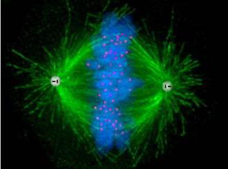
Cellular center (centrosome)
This organelle consists of 2 centrioles and a centrosphere. Centriole cylindrical. Its walls are formed by three microtubules, which merge with each other through cross-linking. Centrioles are arranged in pairs at right angles to each other. It should be noted that cells higher plants lack these organelles.
The main role of the cell center is to ensure uniform distribution of chromosomes during cell division. It is also the center of cytoskeletal organization.
Organelles of movement
Organelles of movement include cilia and flagella. These are miniature growths in the form of hairs. The flagellum contains 20 microtubules. Its base is located in the cytoplasm and is called the basal body. The length of the flagellum is 100 µm or more. Flagella, which are only 10-20 microns, are called cilia. When microtubules slide, cilia and flagella are able to vibrate, causing the cell itself to move. The cytoplasm may contain contractile fibrils, which are called myofibrils - these are organelles of an animal cell. Myofibrils, as a rule, are located in myocytes - muscle tissue cells, as well as in heart cells. They consist of smaller fibers (protofibrils).

It should be noted that myofibril bundles consist of dark fibers - these are anisotropic disks, as well as light areas - these are isotropic disks. The structural unit of the myofibril is the sarcomere. This is the area between the anisotropic and isotropic disk, which has actin and myosin filaments. When they slide, the sarcomere contracts, which leads to the movement of the entire muscle fiber. This uses the energy of ATP and calcium ions.
Protozoa and animal sperm move with the help of flagella. Cilia are the organ of movement of the ciliate-slipper. In animals and humans, they cover the airways and help get rid of small solid particles, such as dust. In addition, there are also pseudopods that provide amoeboid movement and are elements of many unicellular and animal cells (for example, leukocytes).
Most plants cannot move in space. Their movements consist of growth, movement of leaves and changes in the flow of cytoplasm of the cells.
Conclusion
Despite all the diversity of cells, they all have a similar structure and organization. The structure and functions of organelles are characterized by identical properties, ensuring the normal functioning of both an individual cell and the entire organism.
This pattern can be expressed as follows.
Table “Eukaryotic cell organoids”
Organoid | plant cell | animal cell | Main functions |
DNA storage, RNA transcription and protein synthesis |
|||
endoplasmic reticulum | synthesis of proteins, lipids and carbohydrates, accumulation of calcium ions, formation of the Golgi complex |
||
mitochondria | synthesis of ATP, own enzymes and proteins |
||
plastids | participation in photosynthesis, accumulation of starch, lipids, proteins, carotenoids |
||
ribosomes | assembly of the polypeptide chain (protein synthesis) |
||
microtubules and microfilaments | allow the cell to maintain a certain form, are an integral part of the cell center, cilia and flagella, ensure the movement of organelles |
||
lysosomes | digestion of substances inside the cell, destruction of its unnecessary structures, participation in the reorganization of cells, cause autolysis |
||
large central vacuole | provides tension to the cell membrane, accumulates nutrients and waste products of the cell, phytoncides and phytohormones, as well as pigments, and is a reservoir of water |
||
Golgi complex | secretes and accumulates proteins, lipids and carbohydrates, modifies nutrients that enter the cell, and is responsible for the formation of lysosomes |
||
cell center | yes, except higher plants | is the center of organization of the cytoskeleton, ensures uniform divergence of chromosomes during cell division |
|
myofibrils | provide contraction of muscle tissue |
If we draw conclusions, we can say that there are minor differences between animal and plant cells. Wherein functional features and the structure of the organelles (the table above confirms this) has general principle organizations. The cell functions as a coherent and integral system. At the same time, the functions of organelles are interconnected and aimed at optimal functioning and maintaining the vital activity of the cell.